Hydrogen and the MR signal: fMRI from zero, lesson 2
- fMRI
- fMRI Beginner
- MRI Physics
- Neuroimaging
- Data Engineering
Part 2 of 32 · Beginner · fMRI from zero: the series
Every fMRI picture starts as a faint radio signal from the hydrogen in your brain's water. This lesson explains where that signal comes from, in four steps: protons, a magnet, a radio pulse and a coil.
The one-sentence idea
In a strong magnet, hydrogen protons line up a little, and a radio pulse makes them send a signal back.
Explain it simply
The brain is about three quarters water. Each water molecule has two hydrogen atoms, and the centre of each one is a single proton. A proton spins, and that makes it a tiny magnet.
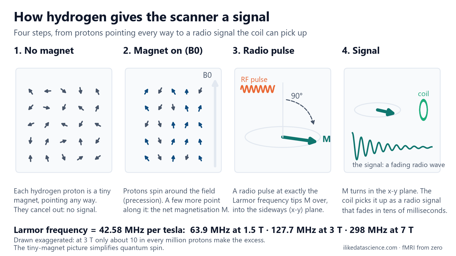
Outside a scanner, these tiny magnets point every way. They cancel out, so there is nothing to measure.
Step 1: the magnet. The scanner's main field, B0, is strong: 3 tesla is about 60,000 times the Earth's field.
In it, the protons wobble around the field direction, like spinning tops. This wobble is called precession.
Step 2: a tiny excess. Slightly more protons point along the field than against it. Their sum is one small magnet, the net magnetisation, written M.
Step 3: the radio pulse. The scanner sends a short radio pulse. It only works at one frequency, the speed of the wobble.
Think of pushing a child on a swing: you must push in time with the swing. The pulse tips M over on its side.
Step 4: the signal. Tipped M keeps turning, like a magnet spinning near a wire.
A receive coil around the head picks this up as a faint radio wave. That wave is the raw MR signal.
For a data engineer, it is like a huge sensor array:
| MRI word | Data engineering view |
|---|---|
| Proton | One tiny sensor with a random reading |
| Net magnetisation | The average of billions of readings: random noise cancels, a small bias remains |
| RF pulse | A broadcast on one exact channel; only listeners on that channel react |
| Receive coil | The antenna that records the reply as a time series |
Go one level deeper
The Larmor frequency. The wobble has a fixed speed, set by the field: 42.58 MHz per tesla for hydrogen.
| Field | Turns per second |
|---|---|
| 1.5 T | about 64 million (63.9 MHz) |
| 3 T | about 128 million (127.7 MHz) |
| 7 T | about 298 million (298 MHz) |
These are radio frequencies, which is why the pulse is called an RF pulse.
How tiny is the excess? At 3 T and body temperature, only about 10 in every million protons make it.
But a 2 mm cube of brain holds about 400 billion billion protons. So the tiny excess still adds up to a magnet the coil can hear.
Why tip it over? M along the field doesn't change, so it makes no signal. Once it turns sideways, its field sweeps past the coil and makes a small voltage.
What happens next. The signal fades within tens of milliseconds, and M slowly grows back along the field.
These two clocks, T2* and T1, are the next lesson. BOLD fMRI reads the signal during that fast fade.
Visual: four steps to a signal
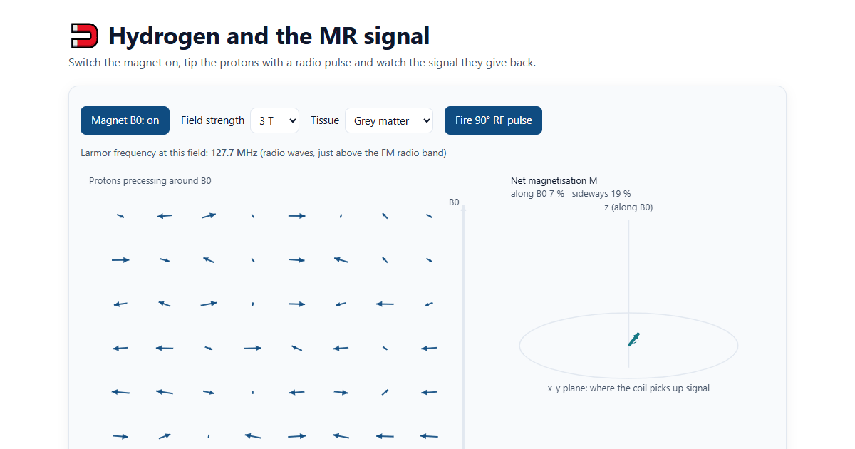
Try it yourself: switch the magnet on, change the field strength and fire the pulse.
Why it matters for data analysis
- The scanner records radio waves, not pictures. Each coil records a time series, and lesson 8 shows how these become images. Most datasets share only the final images (DICOM or NIfTI files).
- Field strength is key metadata. It is stored in the DICOM header and in BIDS as
MagneticFieldStrength. Data from 1.5 T and 3 T scanners differ, so record it and use it when you combine datasets. - Stronger magnets give more signal. The signal grows roughly in step with the field. That is why research uses 3 T and 7 T, but stronger fields also bring more distortion.
- Anything with hydrogen shows up. Water in tissue, blood and the fluid around the brain all give signal. How fast each one fades makes the contrast between them.
Key terms
| Term | Meaning |
|---|---|
| Proton | The centre of a hydrogen atom; it acts like a tiny magnet |
| B0 | The scanner's main magnetic field, measured in tesla (T) |
| Precession | The wobble of a proton around the field direction |
| Larmor frequency | The speed of that wobble: 42.58 MHz per tesla for hydrogen |
| Net magnetisation (M) | The sum of all the tiny magnets in a voxel |
| RF pulse | A short radio pulse at the Larmor frequency that tips M over |
| Receive coil | The antenna around the head that picks up the signal |
Check yourself
- Why does MRI use hydrogen? It is the most common atom in the body, mostly in water and fat. Each hydrogen proton acts as a small magnet, so it gives a strong signal.
- What is the Larmor frequency at 1.5 T? 42.58 × 1.5 = about 63.9 MHz.
- Why must the radio pulse be at exactly the Larmor frequency? Only a pulse in time with the protons' wobble can tip them, like pushing a swing at the right moment. Other frequencies have almost no effect.
Go further
- Hydrogen and the MR signal, the interactive page for this lesson.
- Elster A. D. Questions and Answers in MRI. Short, clear answers on every part of MRI physics.
- McRobbie D. W. et al. (2017). MRI from Picture to Proton, 3rd ed. Cambridge University Press. A friendly book on how MRI works.
For learning only, not medical advice.
Previous: What fMRI is, and what it isn't · Next: Relaxation: T1, T2 and T2*