Electrodes and the 10–20 system: how to read EEG channel names
- EEG
- EEG Beginner
- Electrodes
- Data Engineering
- Neuroscience
Part 6 of 28 · Beginner · EEG from zero: the series
Every EEG file has channel names like Fp1, Cz and O2. They are not random. This lesson shows how electrodes are placed on any head, and how to read a name.
The one-sentence idea
The 10–20 system places electrodes by percentages of head size, so a name means the same spot on every head.
Explain it simply
Think of seat numbers in a stadium. "Block C, row 3" tells you where to sit, whatever the size of the stadium.
The 10–20 system is a naming standard like that, but for heads. Heads differ in size, so it doesn't use centimetres.
It measures each head and places electrodes at 10 % and 20 % steps of those distances. That is where the name comes from.
For a data engineer, it is a shared schema. Two labs that both use it can compare channel C3 with channel C3.
Go one level deeper
How the spots are found. The technician measures from the nasion (the dip between the eyes) to the inion (the bump at the back of the skull).
They also measure from ear to ear. Electrodes go at 10 %, 20 %, 20 %, 20 %, 20 % and 10 % along these lines.
On the front-to-back line this gives:
| Position | Electrode |
|---|---|
| 10 % from the nasion | Fpz |
| 30 % | Fz |
| 50 %, the top of the head | Cz |
| 70 % | Pz |
| 90 % | Oz |
How to read a name. Each name is letters plus a number or a z.
| Part | Meaning | Examples |
|---|---|---|
| Fp | Prefrontal (forehead) | Fp1, Fp2 |
| F | Frontal | F3, Fz, F8 |
| C | Central: the strip across the top | C3, Cz, C4 |
| T | Temporal (sides) | T7, T8 |
| P | Parietal | P3, Pz, P8 |
| O | Occipital (back) | O1, O2 |
| A | Ear lobe, often used as the reference | A1, A2 |
| Odd number | Left side | C3, O1 |
| Even number | Right side | C4, O2 |
| z ("zero") | On the midline | Fz, Cz, Pz |
Numbers also grow away from the midline. C3 sits closer to the middle than T7.
The classic set has 21 positions: 19 on the scalp and 2 on the ear lobes.
Old and new names. For more electrodes, the 10–10 system adds positions halfway between the 10–20 ones. It also renamed four of them for consistency:
| Old name (10–20) | New name (10–10) |
|---|---|
| T3 | T7 |
| T4 | T8 |
| T5 | P7 |
| T6 | P8 |
Both names are still in use, often in the same archive.
Visual: the 10–20 system
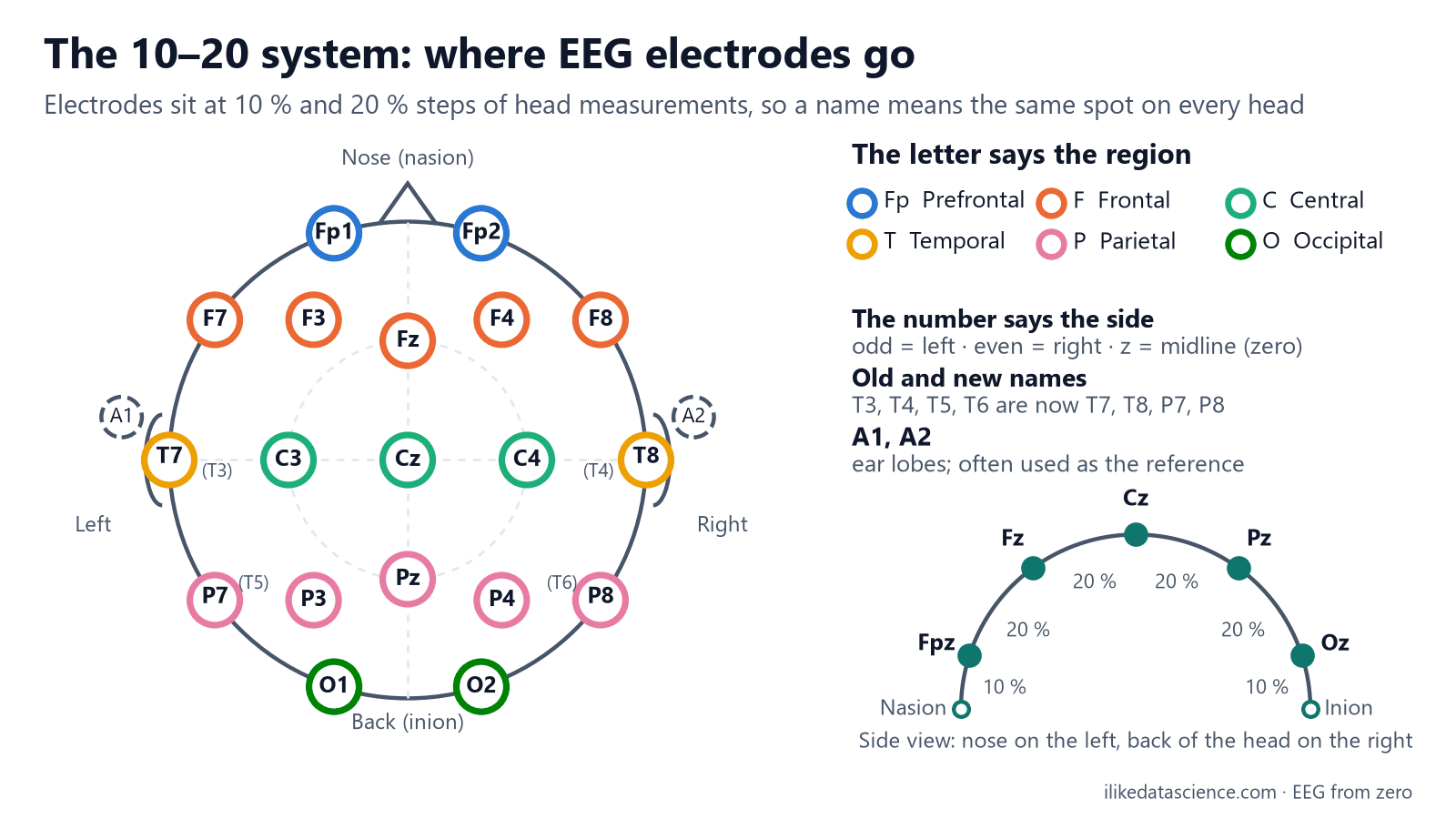
The positions are drawn on an ideal round head, so the picture is exactly even on both sides. Real heads are not.
Why it matters for data analysis
- Channel names are join keys. Merging datasets means matching names. Map the old names to the new ones before you join, or you will lose channels.
- Watch case and spelling. "FP1", "Fp1" and "EEG Fp1-REF" can all mean the same electrode. Clean names into one standard form when you ingest data.
- Positions are data too. Topomaps and source localization need each electrode's position. In MNE-Python, a standard montage (a set of positions) adds them.
- Different devices, different sets. One dataset may have 19 channels and another 64. Pick a shared subset, or use methods that cope with missing channels.
- Name is not region. A name tells you the spot on the scalp, not exactly what lies beneath (lessons 5 and 18).
Key terms
| Term | Meaning |
|---|---|
| 10–20 system | The standard way to place and name EEG electrodes by percentages of head size |
| 10–10 system | A denser version with more positions, and new names for T3 to T6 |
| Nasion | The dip between the forehead and the nose |
| Inion | The bump at the back of the skull |
| Midline | The line from front to back over the top of the head; names end in z |
| Montage (positions) | In MNE-Python, a set of electrode names with their 3D positions |
Check yourself
- Why is it called the 10–20 system? Electrodes go at 10 % and 20 % steps of measured distances across the head.
- Where is C3, and where is O2? C3 is on the left (odd), over the central strip. O2 is on the right (even), at the back.
- An old file has channel T5. What is its newer name? P7. The others are T3→T7, T4→T8 and T6→P8.
Go further
- Acharya J. N. et al. (2016). American Clinical Neurophysiology Society Guideline 2: Guidelines for standard electrode position nomenclature. J Clin Neurophysiol 33(4): 308–311.
- MNE-Python. Working with sensor locations. Standard montages in code.
For learning only, not medical advice.
Previous: The brain's regions and what they do · Next: Noise and artifacts: blinks, muscle, heart, mains hum