Brain waves: delta, theta, alpha, beta and gamma explained
- EEG
- EEG Beginner
- Brain Waves
- Signal Processing
- Neuroscience
Part 4 of 28 · Beginner · EEG from zero: the series
EEG people sort brain rhythms into five bands by speed: delta, theta, alpha, beta and gamma. This lesson gives each band's range, where it is strongest, the state it goes with, and what may be abnormal.
The one-sentence idea
Brain rhythms are grouped by frequency into bands, and each band goes with typical states, places on the head and warning signs.
Explain it simply
Think of the equaliser on a music app. It splits the sound into bass, middle and treble, and shows how loud each part is.
EEG bands do the same for the brain signal. Slow waves are the bass, fast waves the treble.
A frequency is how many times a wave repeats each second, measured in hertz (Hz). A 10 Hz wave goes up and down 10 times a second.
The rule of thumb: the slower the band, the bigger the waves and the deeper the rest. The faster the band, the smaller the waves and the more alert the brain.
Go one level deeper
Band edges follow the international glossary of EEG terms (Kane et al. 2017). Other sources move the edges by about 1 Hz.
| Band | Range | Size | Normal when and where | May point to a problem when… |
|---|---|---|---|---|
| Delta | 0.5–4 Hz | Highest | Deep sleep (N3) in adults; awake in babies and young children | Seen in an awake adult: in one area, a possible lesion; everywhere, a brain-wide problem |
| Theta | 4–8 Hz | Medium | Drowsiness and light sleep; common in children | There is too much in an awake adult (slowing) |
| Alpha | 8–13 Hz | Medium | Relaxed and awake with eyes closed; strongest at the back of the head | The main back rhythm in an adult is slower than 8 Hz |
| Beta | 13–30 Hz | Low | Alert, active thinking; front and centre of the head | There is much more than usual: often a side effect of sedatives such as benzodiazepines |
| Gamma | Above 30 Hz | Lowest | Sensory processing and attention; mostly studied in research | Research only. On the scalp it is easily confused with muscle noise |
Alpha is the classic rhythm. Close your eyes and relax, and alpha appears over the back of the head, near electrodes O1 and O2.
Open your eyes, and it fades. This is one of the easiest EEG effects to see.
Some patterns have their own names. They are not bands, but you will meet them often:
| Pattern | What it is | When you see it |
|---|---|---|
| Mu rhythm | 7–11 Hz arch-shaped waves over C3 and C4, above the movement areas | At rest; it fades when you move, or imagine moving, the opposite hand |
| Sleep spindle | A burst of 11–16 Hz waves lasting at least 0.5 s, strongest over the centre | A sign of N2 sleep |
| K-complex | One big sharp negative wave, then a positive one, lasting at least 0.5 s | A sign of N2 sleep, strongest at the front |
The mu rhythm is the basis of many brain–computer interfaces. Imagining a hand movement changes it, and a model can pick that up.
Visual: the brain wave bands
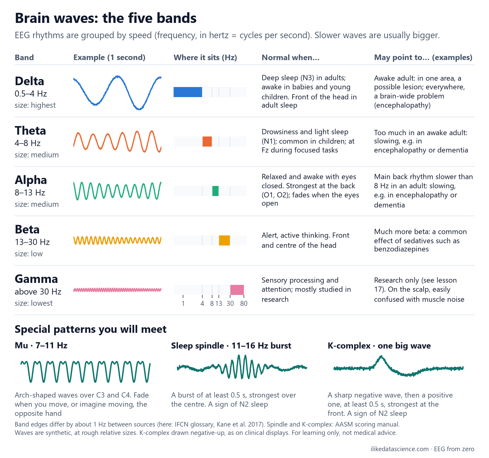
The sample waves are simulated and drawn at rough relative sizes. Delta waves are slow and tall; gamma waves are fast and small.
Why it matters for data analysis
- Band power is the most common EEG feature. You measure how much of the signal falls in each band. Lesson 12 shows how; lesson 22 shows the other features.
- Write your band edges down. Studies use slightly different edges. Store them as settings, not as numbers hidden in code, so results can be compared.
- State changes the bands. Eyes open or closed, drowsiness and age all shift band power. Record them as metadata, or a model may learn the state instead of the disease.
- Drugs change the bands. Sedatives add beta. Ask for medication data before you trust a beta feature.
- Fast bands carry noise. Muscle activity overlaps beta and gamma. Clean first (lessons 7 and 21), then read fast-band power with care.
Key terms
| Term | Meaning |
|---|---|
| Frequency | How many times a wave repeats each second |
| Hertz (Hz) | The unit of frequency: one cycle per second |
| Band | A range of frequencies with a name, such as alpha (8–13 Hz) |
| Amplitude | The size (height) of a wave, in microvolts |
| Slowing | More slow activity than expected for a person's age and state |
| Sleep spindle | A short 11–16 Hz burst that marks N2 sleep |
| K-complex | A large two-part wave that marks N2 sleep |
Check yourself
- When is alpha strongest, and where? Relaxed and awake with eyes closed, at the back of the head (O1, O2). It fades when the eyes open.
- Is delta normal in an awake adult? No. In adults delta is normal in deep sleep (N3). In an awake adult it may point to a problem.
- Why is gamma on the scalp hard to trust? It is small, and muscle activity has a similar frequency. It is easy to mistake one for the other.
Go further
- Kane N. et al. (2017). A revised glossary of terms most commonly used by clinical electroencephalographers. Clinical Neurophysiology Practice 2: 170–185. Band names and edges.
- American Academy of Sleep Medicine. The AASM Manual for the Scoring of Sleep and Associated Events. Spindles and K-complexes.
- Cohen M. X. (2014). Analyzing Neural Time Series Data. MIT Press. Frequency analysis for EEG.
For learning only, not medical advice.
Previous: From brain to file: the signal path · Next: The brain's regions and what they do